·
Non-Destructive Testing Applications
Non-Destructive Testing
(NDT) is a term for describing a class of inspection methods for detecting defects
in objects or structures without destroying the original specimens.
Among the various NDT methods, radiography is one of the popular methods. Radiography uses X-rays and gamma-rays
to produce an image of a specimen on film, showing any changes in thickness, defects
(internal and external), and assembly details to ensure optimum quality in the operation.
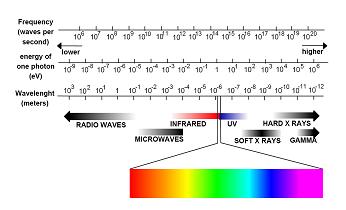
X-rays and Gamma rays are electromagnetic
radiation of exactly the same nature as light, but of much shorter wavelength. Wavelength
of visible light is on the order of 6000 angstroms while the wavelength of x-rays
is in the range of one angstrom and that of gamma rays is 0.0001 angstrom. This
very short wavelength is what gives x-rays and gamma rays their power to penetrate
materials that light cannot. This
high penetration capability of the rays allows us to see the internal conditions
of the object which cannot be seen from outside by naked eyes.
If an x-ray or gamma ray source is placed on one side of a specimen and a
photographic film on the other side, an image is obtained on the film of the thickness
variations in the specimen. This is
a well-established NDT technique and is widely used to detect internal flaws in
weldments and castings and to check for mis-constructions in assemblies
X-rays are produced by an x-ray
generator and is usually described by the electrical voltage across the x-ray tube. The higher the voltage, the greater
is the penetrating power of the radiation; industrial x-ray equipment ranges from
about 20KV to 20 MV and the most powerful equipments can be used to radiograph up
to 500mm steel. Nearly all gamma-radiography
is done with either cobalt-60 or iridium-192 sources.
To obtain good definition images, it is desirable to have small-diameter
radiation sources and the effective source size is in the range of 1 to 4mm diameter. After the radiographic film has been
exposed, it has to be photographically processed (develop, wash, fix and dry) and
is then placed on an illuminated screen for visual interpretation of the image. X-rays and Gamma-rays are
dangerous and must be used either inside a protective enclosure or appropriate barriers
and warning signals.

Site arrangement for carrying out radiography

The above photograph
shows a radiographic test carried out on a butt weld for a welder qualification
test.
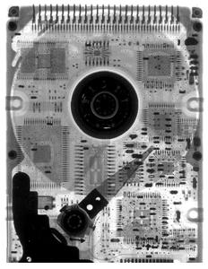
Radiograph of the internal structure of a hard disk
http://www.matsci.ucdavis.edu/MatSciLT/EMS-162L/images/Radiograph.jpg
References:
Ø
http://www.nei.org/howitworks/industrialapplications/
Ø
http://www.geinspectiontechnologies.com/en/products/x-ray/index.html
Ø
http://www.engineershandbook.com/MfgMethods/ndtrt.htm
Ø
http://www.ndt.net/article/mendt03/agfa/agfa.htm
Ø
http://www.ndt-ed.org/EducationResources/HighSchool/Radiography/hs_rad_index.htm
Ø
http://www.ndt-ed.org/EducationResources/CommunityCollege/Radiography/cc_rad_index.htm
Ø
http://www.ndt.net/ndtaz/ndtaz.php